Case Overview
Species: Canine
Gender: Male
Age: 2 years and 5 months
Presenting Complaint: Extensive cutaneous ulceration with myiasis
The patient, “Grey,” sustained severe traumatic lacerations on the dorsum following an attack by three dogs, resulting in large areas of exposed muscle and soft tissue. The dog went missing for several days, during which the wounds became heavily contaminated and infected. Fly eggs hatched within 48 h, producing extensive myiasis with maggot infestation that rapidly destroyed necrotic tissue. The lesions progressed to involve the dorsal, perineal, genital, and abdominal regions, presenting as multiple honeycomb-like ulcers. The owner subsequently transported the dog to a local veterinary hospital for emergency care.
Initial Management
Upon admission, the veterinary team performed complete clipping of the coat and manual removal of maggots with forceps. This was followed by systematic wound irrigation with ozonized water to debride necrotic tissue and disrupt microbial biofilms.
Treatment Protocol
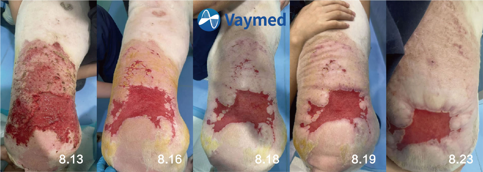
Beginning on August 13, sequential therapy was instituted as follows:
1. Ozonized Water Irrigation
Daily systemic wound lavage for antimicrobial action, biofilm disruption, and inflammatory modulation.
2. Class IV Laser Therapy
Performed immediately after lavage, using the ‘Dog–Skin–Wound’ mode, once daily, for 7 consecutive days.
Application method: maximal scanning area, treated separately on the left and right sides; irradiation performed with uniform Z-shaped or S-shaped sweeping movements.
Duration: approximately 12 minutes per session.
After 7 days of continuous treatment, the wound surface showed marked reduction in size, with robust granulation tissue formation. At the time of reporting, the wound was nearly healed.
Potential Risks and Complications of Extensive Ulceration
· Local infection: Bacterial (e.g., Staphylococcus, Streptococcus, Pseudomonas) or fungal invasion may lead to abscess or sinus tract formation within 48 h; deep infections may extend to periosteum or joints, resulting in osteomyelitis or septic arthritis.
· Sepsis/septicemia: Bacterial dissemination and toxin release can induce systemic inflammatory response syndrome (SIRS). Reported mortality in canine and feline sepsis is approximately 20–40%.
· Necrosis and gangrene: Tissue ischemia compounded by infection may lead to progressive necrosis. Animals with diabetes, advanced age, or immunosuppression are at higher risk, sometimes necessitating amputation.
· Chronic ulceration: Persistent infection, poor nutrition, or continuous pressure (e.g., sternum, hip tuberosity) may delay healing for weeks to months, forming refractory ulcers.
· Joint contracture and functional impairment: Involvement of tendons or articular structures may lead to fibrotic contracture, resulting in impaired ambulation, mastication, or defecation.
Home care precautions prior to veterinary consultation: Avoid the use of alcohol or hydrogen peroxide for wound cleansing to prevent damage to granulation tissue; prevent the patient from licking or biting wounds; restrict activity; and avoid water exposure to minimize secondary infection risk.
Summary
Ozonated water creates a “chemical debridement” environment that provides rapid bactericidal action, disrupts biofilms, and modulates inflammation. Class IV laser therapy promotes “physical–biochemical” accelerated repair by inducing photobiomodulation, enhancing microcirculation, and stimulating collagen remodeling. Sequential application of these two modalities can reduce the healing time of chronic ulcers by 25–30%, increase the complete healing rate by more than 40%, and significantly lower the risk of secondary infection and hypertrophic scarring.
Contact Information
Phone Number:+86-027-81808016
Email:info@vaymed.com
Address:3rd Floor, Building #32-2, No.6 Hejiahu Street, Jiangxia District, Wuhan, Hubei, PR.China